
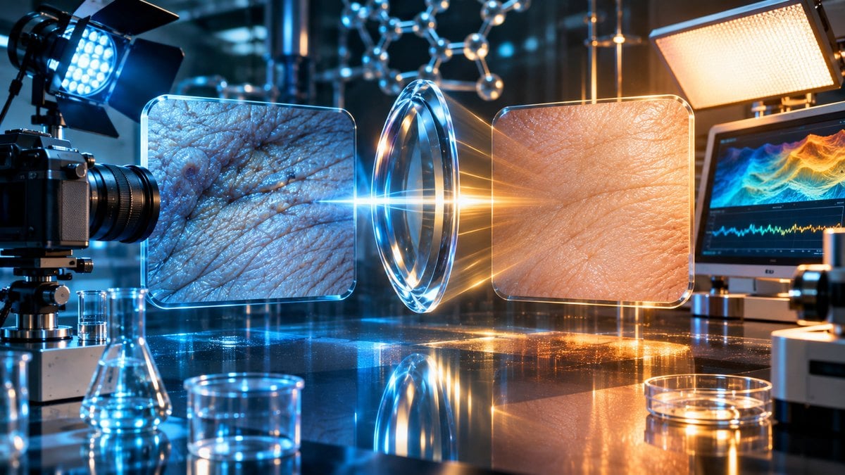
Two photographs appear side by side in a cosmetic research presentation. In the first, the subject’s skin looks uneven, with visible fine lines and pronounced shadows. In the second, the same area appears smoother and brighter. The images invite an immediate conclusion: an experimental ingredient must have improved the skin.
But imagine discovering that the second photograph was captured using softer lighting, a slightly different camera angle and a different facial expression. The apparent improvement might partly reflect the photography rather than a measurable change in the skin.
That possibility is particularly important when scientific findings are communicated through before-and-after photographs. Images can illustrate research results, but they can also introduce uncertainty when acquisition conditions are inconsistent.
For researchers investigating cosmetic peptides, collagen-related measurements and skin appearance, the central question is not simply whether the second photograph looks better. It is whether the study used reliable methods to establish what actually changed.
Why photographs can make a cosmetic result look more convincing than the evidence
Human vision is exceptionally responsive to differences in brightness, contrast, texture and facial appearance. A side-by-side photograph can therefore make a visual comparison feel persuasive before the viewer has considered how the images were produced.
Clinical and research photography attempts to reduce this problem through standardization. Investigators try to maintain consistent photographic conditions so that differences between images are more likely to reflect changes in the subject rather than changes in the equipment or environment.
var infolinks_wsid = 36;
A systematic review published in JAMA Dermatology in 2015 examined technical and procedural standards for camera-acquired dermatological images. Its authors identified important variables including lighting, background, camera position, spatial resolution, image processing and color calibration.
The review also found that universally accepted imaging standards had not been established across the field at that time.
That conclusion matters because a photograph’s apparent clarity is not the same as its scientific comparability. Two visually attractive images may have been acquired under conditions that make meaningful interpretation difficult.
A strong cosmetic study needs more than professional-looking photography. It must establish whether its imaging methods were sufficiently consistent for the research question being investigated.
How lighting can hide or exaggerate fine lines
Lighting changes the way surface texture appears in a photograph. A directional light source may create pronounced shadows around small changes in skin contour. More diffuse lighting can reduce those shadows and make the same surface appear smoother.
This does not necessarily mean that the structure of the skin has changed.
The camera is recording reflected light, and the appearance of surface irregularities depends partly on how that light reaches the subject and returns to the imaging system.
A photograph taken under strong directional illumination may emphasize fine lines that appear less conspicuous when the same area is photographed under softer illumination.
The 2026 review “Photography in Dermatology,” published in Frontiers in Medicine, emphasizes consistent lighting, camera positioning and photographic conditions when documenting skin characteristics.
For repeated photographs, the review recommends maintaining a consistent location and orientation relative to the light source whenever possible.
This principle is important in studies involving claims about skin texture. If the lighting changes between baseline and follow-up photographs, the images may not provide reliable evidence of the magnitude of any actual change.
Researchers must distinguish an improvement in photographic appearance from an independently measured improvement in the biological or physical property being investigated.
Why a small camera-angle difference can change the apparent outcome
Camera position influences perspective. The apparent proportions and contours of a photographed surface can change when the camera moves closer, farther away, above or below the original viewpoint.
This becomes especially important when researchers compare small features such as facial lines, surface texture or local skin contours.
A slight change in head position may alter the shadows around the eyes, mouth or forehead. Differences in camera height can also affect the apparent shape of the photographed area.
A 2017 article in the International Journal of Dermatology, “Standardized Methods for Photography in Procedural Dermatology Using Simple Equipment,” describes the importance of consistent photographic settings, patient position and framing.
These methods are relevant to cosmetic research because a visual comparison is meaningful only when the images document reasonably comparable views.
A photograph taken from an advantageous angle is not necessarily false. However, when the comparison photograph uses a different angle, the pair may exaggerate or obscure a change.
Researchers should report their photographic procedures rather than expect readers to infer standardization from the images alone.
The overlooked influence of facial expression and skin tension
Facial expression changes the appearance of the skin. Smiling, squinting, raising the eyebrows or relaxing facial muscles can alter the visibility of certain lines and folds.
This creates another challenge for before-and-after comparisons.
A baseline photograph showing an animated expression may display different contours from a follow-up photograph showing a neutral expression. The difference can make a region appear smoother even without demonstrating a lasting structural change.
Consistency in subject positioning and expression is therefore relevant when the measured outcome involves facial appearance.
Scientific photography should also provide enough context for readers to understand which anatomical region is being evaluated.
A tightly cropped image may direct attention to a small area while concealing differences in head position or camera orientation that would be apparent in a wider view.
For reliable interpretation, photographs should be considered alongside the study’s documented methods and other relevant measurements.
What image processing can change after a photograph is captured
Modern cameras do not always produce a simple, untouched representation of the light entering the lens.
Digital imaging systems may apply automatic processing involving exposure, contrast, sharpening, noise reduction and color balance.
Smartphones can use computational photography techniques that combine information from multiple exposures or adjust local characteristics of the image.
Some processing improves visibility and photographic quality. However, inconsistent processing can complicate comparisons when the research outcome depends on subtle differences in color or surface texture.
The 2026 Frontiers in Medicine review notes that smartphone image-processing algorithms may affect the fidelity of dermatological photographs by changing clinically relevant color and surface characteristics.
This matters when studies rely on photographs to communicate changes in redness, pigmentation or fine lines.
If one photograph receives different processing from another, the comparison may contain a technical difference unrelated to the experimental ingredient.
Image acquisition and processing procedures should therefore be sufficiently documented for researchers to evaluate their consistency.
Why visible smoothness is not the same as increased collagen
Skin appearance is influenced by multiple structural and environmental factors. The visual prominence of surface lines may depend on hydration, lighting, expression and the arrangement of tissues beneath the surface.
Collagen is a family of structural proteins involved in the organization of connective tissue. But the apparent smoothness of a photograph is not a direct measurement of collagen abundance or organization.
A claim about collagen production requires evidence appropriate to that biological process.
Photographs may show whether a visible feature appeared different under specified conditions. They cannot, by themselves, demonstrate which molecular process caused that appearance.
This distinction is particularly relevant when cosmetic products are described using scientific language about collagen, peptides or cellular signaling.
A plausible mechanism does not establish that a visible result occurred for the proposed reason.
Readers exploring interpreting collagen research should examine what the investigators measured, whether the outcome was independently assessed and whether the conclusions were supported by the reported data.
Changes in photography, skin hydration and tissue biology should not be treated as interchangeable outcomes.
What skin hydration instruments measure that photographs cannot
Skin hydration can be investigated using instruments that assess properties associated with water in the outermost layer of the skin, known as the stratum corneum.
Some devices use electrical capacitance or related measurement principles to estimate hydration-associated characteristics.
These measurements provide information that is different from the visual appearance captured in a photograph.
However, instrument readings also have limitations. Environmental conditions, measurement site, instrument performance and participant-related variables may affect the result.
International guidelines published in Skin Research and Technology in 2013 address the measurement of skin hydration and transepidermal water loss, emphasizing the need to minimize or recognize sources of variability.
The guidelines discuss environmental conditions, participant acclimatization, instrumentation and the importance of consistent reporting.
They demonstrate why an instrument-generated number should not be assumed reliable simply because it appears objective.
Both photographic and instrument-based methods require appropriate controls. The difference is that each method measures a different property and introduces its own potential sources of error.
Why transepidermal water loss is not a photograph of skin health
Transepidermal water loss, or TEWL, concerns the movement of water vapor through the skin barrier into the surrounding environment.
Researchers use TEWL measurements as one way of investigating aspects of skin barrier function.
A TEWL instrument does not produce the same information as a photograph showing the surface appearance of the skin.
Nor is TEWL identical to a hydration measurement. Skin hydration concerns water-associated properties within the stratum corneum, while TEWL concerns outward water-vapor movement.
The European Expert Group on Efficacy Measurement of Cosmetics, commonly known as EEMCO, has published guidance emphasizing the importance of standardized conditions when measuring TEWL.
Environmental temperature, humidity, airflow and other variables can influence readings.
Therefore, a study reporting lower TEWL should describe how the measurements were obtained and interpreted. It should not automatically translate a change in one instrumental outcome into a broad claim about appearance or clinical benefit.
Similarly, a photograph that appears smoother does not prove that skin barrier function improved.
Different outcomes require different supporting evidence.
Why the environment can influence the apparent result
Scientific measurements do not occur in isolation from their surroundings.
Skin-related measurements may be influenced by room temperature, relative humidity and the participant’s recent environment. Photography can also change with ambient light and the properties of the surrounding room.
This means environmental consistency is important for both visual documentation and instrument-based testing.
The international skin-property measurement guidelines emphasize accounting for environmental and participant-related sources of variation, including acclimatization to the measurement environment.
When those conditions differ substantially between assessments, the resulting data may become harder to compare.
An apparent before-and-after improvement may still be genuine, but researchers need to investigate whether an alternative explanation could account for some of the measured difference.
The strongest study designs attempt to reduce avoidable variation and transparently report the conditions under which measurements occurred.
Where cosmetic peptide research fits into the evidence
Peptides are molecules composed of amino acids connected through peptide bonds. Their biological roles vary widely, and experimental studies investigate different compounds for different scientific purposes.
Some cosmetic research examines specific peptide-containing formulations and their relationships with skin-related measurements.
One frequently discussed compound is the copper-associated tripeptide GHK-Cu. Its research history includes laboratory investigations and comparatively small human studies of topical cosmetic formulations.
The American Academy of Dermatology notes that human evidence concerning copper-peptide skin claims remains preliminary, despite some encouraging findings in small trials.
Those findings should not be generalized to every peptide or every product described using similar terminology.
Formulation characteristics, study methods and the precise compound investigated can all affect the interpretation of results.
Educational material on copper peptide laboratory studies should distinguish molecular mechanisms, preclinical observations and controlled human outcomes.
It should also distinguish a photograph illustrating an observation from evidence establishing the cause of that observation.
Scientific accuracy requires identifying the intervention, the research model and the outcome measured rather than assuming that all peptide-related findings support a single conclusion.
How small cosmetic studies can amplify uncertainty
Small studies can be useful for exploring new questions, but they often provide less precise estimates than larger, well-designed investigations.
Participants may differ in baseline skin characteristics, environmental exposures and other variables related to the measured outcome.
When only a small number of participants are included, a few unusually large or small responses can substantially influence the average result.
The challenge becomes greater when researchers investigate many outcomes without clearly distinguishing primary measurements from exploratory analyses.
A study may examine hydration, elasticity, appearance, participant satisfaction and multiple photographic ratings. Finding a favorable result among several measurements does not automatically establish that the treatment produced a broad benefit.
Readers should examine the study design, comparison group, effect size and uncertainty around the estimated result.
Research communication becomes misleading when a single visually dramatic image receives more attention than the complete pattern of findings.
Photographs are especially memorable. Their persuasive effect should not be allowed to substitute for the strength of the underlying study.
Why blinded assessment matters when appearance is the outcome
Human observers can be influenced by expectations, even when they intend to judge images carefully.
If an evaluator knows which photograph represents the follow-up condition, that information may affect how subtle changes are interpreted.
Blinded assessment can help reduce this concern when it is feasible and appropriate for the research design.
Researchers may also use structured rating scales or other predefined evaluation methods to improve consistency.
However, a rating scale is only useful when it has appropriate measurement properties for its intended purpose.
Blinding does not eliminate problems caused by inconsistent photography. An evaluator may remain unaware of treatment assignment while still seeing images taken under different lighting conditions.
The quality of image acquisition and the independence of assessment therefore address different potential sources of bias.
Strong research methodology considers both.
How readers can evaluate an impressive before-and-after comparison
The first question is whether the images appear to represent comparable views.
Differences in framing, camera position, facial expression or lighting may affect the appearance of skin features independently of any biological change.
The second question concerns the study’s measurements. Was the conclusion based entirely on a visual judgment, or did investigators use additional methods relevant to the claimed outcome?
The third question concerns the comparison group. Without an appropriate comparison, it can be difficult to distinguish an experimental effect from other changes occurring during the study.
The fourth question is whether the results have been independently reproduced.
A single promising study may justify further investigation without establishing a reliable general conclusion.
Readers using this local research guide as a starting point for understanding peptide-related science should separate research terminology from the actual strength of the evidence.
A product description, scientific hypothesis or attractive photograph does not establish an approved clinical use.
The same caution applies to substances described as experimental or research-grade. Chemical identification and analytical purity are separate from evidence of human safety or effectiveness.
What more convincing skin research would look like
A well-designed study begins with a clearly defined question and an outcome that can meaningfully address it.
If researchers intend to examine skin hydration, they should use methods suited to that property. If their objective concerns visible lines, they should define how those features will be measured and how photographic consistency will be assessed.
When investigating biological changes involving collagen or cellular signaling, researchers need evidence capable of addressing those mechanisms rather than relying solely on visual appearance.
Appropriate comparison groups, predefined outcomes and transparent reporting improve the interpretability of findings.
Photographs can remain valuable supporting material. They help readers visualize the observed features and may document changes that are difficult to describe in words.
But photographs become stronger scientific evidence when their acquisition conditions are controlled and their interpretation is supported by appropriate measurements.
Independent replication further strengthens confidence by showing whether comparable results occur outside the original study conditions.
Responsible interpretation leaves room for uncertainty rather than turning every favorable observation into a definitive statement.
The important difference between looking better and being proven better
Photography can reveal genuine changes in appearance, but it can also magnify differences caused by illumination, perspective, expression and digital processing.
This makes standardized imaging especially important when cosmetic research depends on subtle visual features.
Peptide-related investigations add another layer of complexity because molecular mechanisms, laboratory responses and human outcomes are not interchangeable forms of evidence.
Studies assessing skin appearance may use photographs, hydration instruments, barrier-function measurements and other methods. Each contributes information, but none should be credited with proving more than it actually measures.
The most persuasive cosmetic research is not necessarily the study with the most dramatic before-and-after picture. It is the study that defines its outcomes clearly, controls relevant sources of variation and reports results in a way that other investigators can evaluate.
A photograph can begin an interesting scientific conversation. Reliable measurements, transparent methods and independent evidence determine how much of the apparent change can reasonably be believed.